Taxonomic research required taxa, having morphological variations caused by frequent hybrid and/or vegetative reproduction in nature, have been called by several categories; microspecies (Davis and Heywood, 1963), superspecies (Mayr, 1969), semispecies (Grant, 1957; Baum, 1972), multispecies (Van Valen, 1976), species complex (Grant, 1981) etc. The Viola albida complex, consists of three taxa, V. albida var. albida, V. albida var. takahashii, and V. albida var. chaerophylloides, and these are sympatric (Kim et al., 1991). The frequent hybridization among taxa in this complex takes places, and there are almost absence of differences in taxonomic features for dividing species except for various leaves shape (Russell, 1960; Kim, 1986; Kim et al., 1991).
It has generally known that V. albida var. takahashii might be originated by the hybridization between V. albida var. albida and V. albida var. chaerophylloides (e.g., Kim, 1986). There is, however, no direct evidences regarding this so far. This complex shows numerous kinds of leaf forms in-between simple to palmately compound leaf, so that it is taxonomically difficult to explain (Kim and Lee, 1988; Kim et al., 1991; Jang et al., 2006; Whang, 2006). The results of morphological researches about this complex should give rise to debate in both the selection of scientific name (Regel, 1861; Maximowicz, 1877; Becker, 1902; Makino, 1912; Nakai, 1922a, 1922b; Ishidoya, 1929; Maekawa, 1954; Chung, 1959; Ito, 1962; Hashimoto, 1967; Lee, 1969, 1980; Park, 1974; Kim et al., 1991; Lee, 2003) and their classification system (Becker, 1925; Takenouchi, 1955; Maekawa and Hashimoto, 1963; Fu and Teng, 1977; Kim, 1986; Jang, 2012). Recent researches based on molecular data like DNA sequences, RAPD, ISSR, etc have supported the design adequacy of this complex (Yoo et al., 2004, 2005; Whang, 2006; Yoo and Kim, 2006; Koo et al., 2010). Further researches, however, are required to have better understand at the speciation and/or evolution in this complex. We have been conducting four different experiments on this complex; developmental investigation on early leaf morphogenesis (Choi, 2011), cross test between V. albida var. albida and V. albida var. chaerophylloides in both laboratory and wildness (An, 2015), and both cloning and expression of several key genes responsible for leaf morphogenesis (Srikanth, 2014; Srikanth et al., 2019).
This study aims to determine morphological segments appearing in the early leaf development of Viola albida complex in order to give developmental clue for distinguishing taxa in the complex. The new data would be also useful as a basic reference for genes isolation responsible for different leaf morphogenesis if there are some successful results.
Materials and Methods
Plant materials
More than 1,000 individuals in Viola albida complex were collected from 2005 to 2015 at Naejangsan and Jeoksangsan Mountains for this study. They were transplanted at laboratory of about 66 m2 that has been regulated in both temperature and intensity of illumination, and their growth and development were observed more than seven years. The facilities locate in The 1st Science Building of Chonbuk National University. The individuals showing constant in shape during the several generations were selected for developmental studies.
Seed germination and collection of young shoots
The matured seeds were collected from both chasmogamous and cleistogamous flowers, and then stored at 4°C refrigerator. They were washed in tap water with liquid detergent for five minutes, and then sterilized using 70% ethanol and a mixture of distilled water (250 μL), lax (250 μL), and Triton X-100 (25 μL). Sterilization was done by dipping the seeds in ethanol 70% for 1 min and transferring them to sodium hypochloride solution at 2% for 20 min. Sterilized seeds were inoculated into 150 mL glass flasks containing 50 mL of MS medium (Murashige and Skoog, 1962) supplemented with 0.4 mg/L thiamine, 1 mg/L pyridoxine, 0.5 mg/L nicotinic acid, 100 mg/L mio-inositol and 0.5 g/L hydrolyzed casein, diluted for the concentration defined in each treatment. The inoculation process was done under aseptic incubator in a sterile environment. The flask inoculated with sterilized seed were capped with plastic lids and transferred to a growth room. They were remained for six months at 23°C with 16 h lighting at an intensity of eight foot candles. Seeds were monitored daily and were evaluated for contamination and seed germination.
Scanning electron microscopy
Young shoots serially collected were fixed by 4% glutaraldehyde solution for the purpose of scanning electron microscopy (SEM) observation. After full fixation, young shoots were washed with distilled water and dehydrated with a graded ethanol series (30, 50, 70, 80, 90, 95, and 100%) consisting of 10 min steps for each ethanol concentration followed by a graded ethanol-acetone series with three steps. Young shoots were subsequently freeze dried (VFD-21S, Hidachi, Tokyo, Japan), mounted on SEM stubs with double-sided adhesive tape, coated with gold in 15 nm thickness, and examined with a Hidachi ISI ABT (SR-50) SEM operated at 10 kV. All the stubs prepared are housed in the Botany Laboratory of Chonbuk National University.
Results
General features in early leaf development
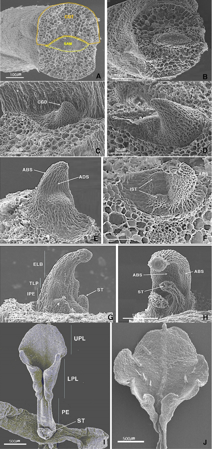
The shape of shoot apical meristem (SAM) before germination is long elliptical in all taxa of Viola albida complex (Fig. 1A, B). The SAM, constituted with about 200 cells, of V. albida var. albida is a little bit smaller than those of V. albida var. takahashii and V. albida var. chaerophylloides, about 250 cells. After germination, the SAM showed gradual changes to develop young leaf. The morphogenesis of young leaves then were rapidly made up within a week, and their size reached up to around 1,000 μm in width and 3,000 μm in length (Figs. 1–3). The morphological segments of early leaf development, appeared in the taxa of the complex, could be distinguished in comparing the features of both mature leaves as well as general shapes in very early stages of germination. Morphological segments of early leaves development detected in this study were different among taxa in the complex (Figs. 1–3).
Morphological segments of early leaves in Viola albida complex
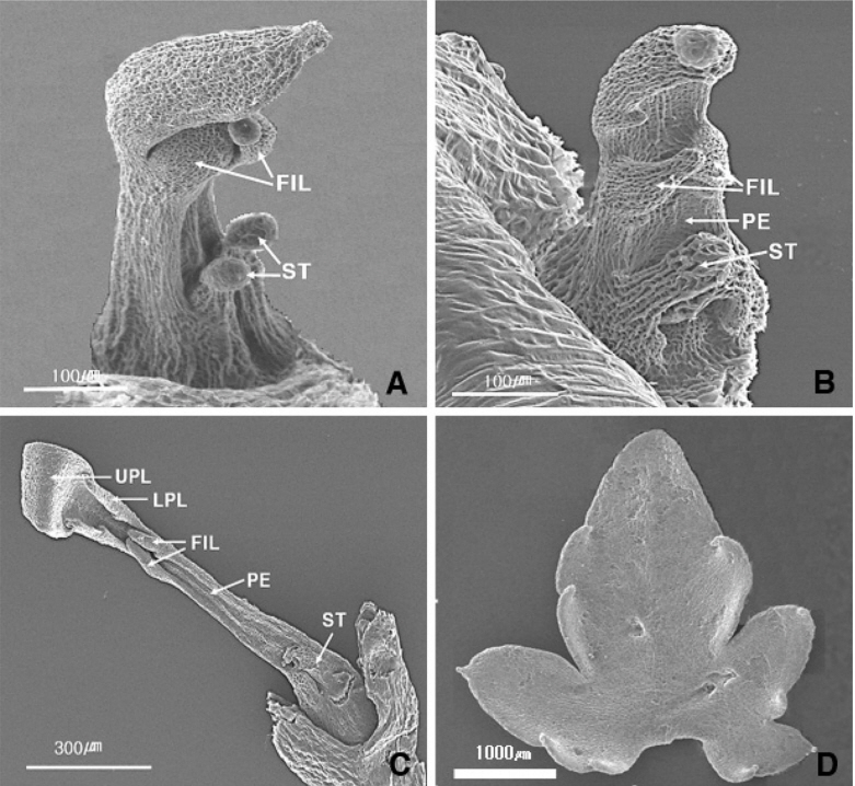
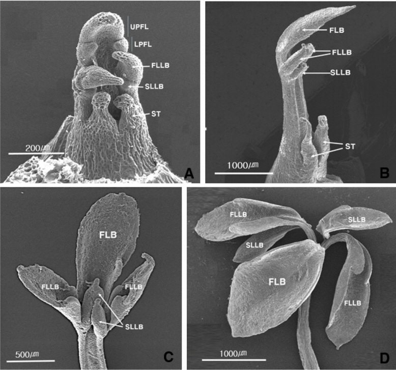
The features of early leaf development in V. albida var. albida was showed in Fig. 1 in detail. There are the SAM before germination (Fig. 1A), the initiation of germination (Fig. 1B), the initiation of conical growth directionally (Fig. 1C), ended up conical growth (Fig. 1D), the formation of adaxial and abaxial side of leaf (Fig. 1E), the initiation of stipule (Fig. 1F), the formation of transitional zone between leaf and petiole (Fig. 1G), the expansion of upper part of leaf (Fig. 1H), the formation of almost all parts of early leaf (Fig. 1I), and the adaxial side of early simple leaf (Fig. 1J). The features of early leaf development in V. albida var. takahashii was showed in Fics 1–2 in detail. Its morphological segments during early leaf development are the same as those of V. albida var. albida till the formation of transitional zone between leaf and petiole. The different features are, however, the first initiation of lateral lobe in both parts of leaf (Fig. 2A), the expansion of leaf blade (Fig. 2B), the formation of almost all of early leaf (Fig. 2C), and the adaxial side of early lobed simple leaf (Fig. 2D). The early leaf development of V. albida var. chaerophylloides was showed in Fics 1–3 in detail. Its morphological segments during early leaf development are the same as those of both V. albida var. albida till the formation of transitional zone between leaf and petiole and V. albida var. takahashii till the first initiation of lateral lobe of the leaf. The different features are, however, the first initiation of second lateral lobe in both parts of leaf (Fig. 3A), the formation of almost all of early leaf (Fig. 3B), the expansion of leaf blade (Fig. 3C), and the early palmately compound leaf (Fig. 3C).
In conclusion, taxa within the complex could be distinguished based on the features described above. The morphological segments of early leaf development in V. albida var. albida were categorized into eight stages: I, the initiation of shoot germination; II, the conical growth directionally of leaf; III, the adaxial and abaxial formation of leaf; IV, the initiation of stipule; V, the formation of transitional zone between leaf blade and petiole; VI, the expansion of upper part of leaf blade; VII, the formation of almost all part of early leaf; VIII, the early palmate compound leaf. Viola albida var. takahashii is clearly separated from V. albida var. albida by the features of both V-1, the initiation of first lateral lobe at both parts of early leaf, and VIII, the early lobed simple leaf. Viola albida var. chaerophylloides is also separated from V. albida var. albida and V. albida var. takahashii by three features, V-1, the initiation of first lateral lobe at both parts of early leaf, V-2, the second lateral lobe at the below of first lateral lobe, and VIII, the early palmate compound leaf.
Discussion
Viola albida var. albida distributes widely on the Korean Peninsula, and its speciation course is of interest. This species complex consists of three taxa, V. albida var. albida, V. albida var. takahashiii, and V. albida var. chaerophylloides (Kim et al., 1991). Interestingly, it is not difficult to find the spots where all taxa in this complex are sympatric distribution or neighborhood areas across many of mountains (Whang, 2006). Taxa in this complex hardly find differences in taxonomic characters except for various leaves shapes. There are, however, numerous leaf shapes from simple to palmately compound forms together with intermediate types in-between, so that the establishment of both species limitation and classification have long been debating among researchers (e.g., Kim et al., 1991; Whang, 2006; Jang, 2012). Many researches on this complex have been conducted because of this exciting attention to taxonomists (e.g., Kim et al., 1991; Jang, 2012). The authors have been also studying for several decades to this complex in order to find the courses of speciation and evolution (Whang and Kim, 1985; Kim et al., 1991; Whang, 2006; Koo et al., 2010).
It is too difficult to prove the evolution of organism that is a phenomenon gradually taking place for a long time (e.g., Barrick et al., 2009). It is also never easy for what the experimental demonstration as to morphological changes taking place in real time on the basis of the evolutionary point of view. In these context, we have recently done four different experiments for this complex: 1) the investigation of morphogenesis from seed germination to early leaf shape (Choi, 2011), 2) the investigation of the leaf features of second generation after the cross test in both laboratory and wildness (An, 2015), 3) cloning of genes that have directly related to the morphological segments during early leaf development (Srikanth, 2014), and 4) and the investigation of the expression pattern of their genes (Srikanth, 2014; Srikanth et al., 2019).
This article is about a comparative study of early leaf development showing the morphological segments. This data, showing developmental differences, were primary useful as some diagnostic features to distinguish the taxa in the complex. This data would be employed not only for cloning of genes that have directly related to the morphological segment during early leaf development, but also for testing their differential expression pattern (Srikanth et al., 2019).
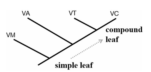
The data obtained this study were Figs. 1–3, and their scientific interpretations summed up in Fig. 4 and Table 1. On the basis of Table 1, the crucial morphological differences among taxa could be summarized as follows. After the development of feature V (the formation of transitional zone between leaf blade and petiole) in V. albida var. albida, two features, V-1 (the initial of first lateral lobe at both parts of early leaf) and V-2 (the initial of second lateral lobe at the below of first lateral lobe), were repeatedly developed in V. albida var. takahashii and V. albida var. chaerophylloides respectively. There were, therefore, absent in two features like V-1 and V-2 in V. albida var. albida, and also absent in the feature V-2 in V. albida var. takahashii, so that these two taxa were clearly distinct from V. albida var. chaerophylloides. In addition, two different types of heterochrony, like spatial and temporal changes in development in a descendant relative to its ancestor, has long been known as the mechanisms most important for evolution (Haeckel, 1875).
The meaning of above differences would be that two taxa, V. albida var. takahashii and V. albida var. chaerophylloides, are peramorphosis comparing to the ancestor, V. albida var. albida. Because these two taxa have evolved excessive stages besides all developmental stages of ancestor type (e.g., Buendía-Monreal and Gillmor, 2018). A suggested topology based on the above shows in Fig. 4. In putting V. mandshurica having simple leaf as an out-group, V. albida var. albida had initially originated in the Korean Peninsular and/or its neighborhood area, and then V. albida var. takahashii and V. albida var. chaerophylloides might have been originated caused by hybridization with other species and/or genetic changes.


